Conservation Journal
Spring 2002 Issue 40
Further applications of Raman microscopy in paper conservation
Introduction
Raman microscopy is becoming increasingly popular as an analytical technique in the scientific and conservation departments of museums. The power of the technique stems from its ability to permit the unambiguous identification of particles of micrometre dimensions both non-destructively and in situ. Raman can be readily applied as an investigative tool in a variety of situations. A previous article (V&A Conservation Journal, Vol 30 1999) looked at the use of Raman for pigment analysis on different types of objects. The versatility of Raman was illustrated with successful analysis being carried out on a range of objects from portrait miniatures to oil paintings and also three-dimensional sculpture. Furthermore, with the portrait miniature, it was possible to carry out the analysis through the cover glass. The theory of Raman spectroscopy is described in the literature. In practice a low-powered laser light is directed onto the material to be analysed. The difference in energy between the original and scattered photons provides the Raman spectrum, which is a ‘fingerprint‘ of the molecule under investigation. Identification of the material is then made by reference to a library of spectra.
Identification of Salt Crystals in European Portrait Miniatures
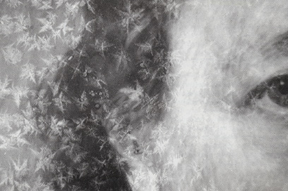
Figure 1. Miniature on ivory showing crystal formation (Museum no. P.22-1928). Photography by Alan Derbyshire (click image for larger version)
Three portrait miniatures on ivory were analysed by Raman microscopy to determine the identity of tiny, white crystals, which had formed under, within, or on top of their paint layers. Some crystals also formed on the verso of the ivory support away from any paint layer. In each case the crystals were identified as magnesium hydrogen phosphate trihydrate, also known as newberyite (MgHPO4.3H2O). Small, white crystals found growing on the inner surface of ivory tusks were also identified as newberyite by means of Raman microscopy. The authenticity of a sample of magnesium hydrogen phosphate trihydrate was checked by powder X-ray diffraction. Thus, it is concluded that the tiny, white crystals occurring on the portrait miniatures on ivory almost certainly originate from the ivory substrate – probably under conditions of high humidity.
Investigation of the effect of iron gall ink on the discoloration of lead white pigment
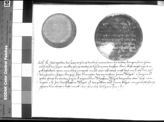
Figure 2. Jamnitzer Manuscript (NAL), p48, showing example of ghost image running through lead white. Photography by Alan Derbyshire (click image for larger version)
This research stemmed from a recently observed phenomenon in a manuscript from the sixteenth century. The manuscript is by Wenzel Jamnitzer, a German goldsmith. It has been observed that, within the book, large areas of the lead white pigment have blackened. However, areas of the lead white which correspond directly to iron gall ink inscriptions on the reverse of the same sheet are not blackened. Effectively they remained white; i.e. a ghost image of the inscriptions can be seen running through the otherwise discoloured lead white pigment (figure 2).
To begin to understand this effect it was necessary to be able to carry out non-destructive, non-invasive analysis of a number of the pigments and discoloured pigments involved. This was successfully completed using Raman microscopy and X-ray fluorescence spectroscopy (XRF). The analysis confirmed the presence of lead white and other pigments including the little-known pigment, mosaic gold (tin disulphide). A Raman spectrum for blackened lead white i.e. lead sulphide was also obtained, in situ, for the first time. Having confirmed the presence of iron gall ink and various pigments and their degradation products it was possible to suggest possible mechanisms for the ghost image effect. Observations on the possible source of the lead white discoloration were also made.
Identification of pigments in Indian miniatures – The Hamzanama
Raman spectroscopy was also used to identify pigments from the Hamzanama, a painted manuscript from the Mughal period (1526-1707). This was a joint project with the Christopher Ingold laboratories at University College London (UCL). The non-destructive and non-invasive nature of this analytical technique was especially attractive. Also the portability of the Raman microscope allowed analysis to be conducted on site.
The Victoria and Albert Museum has 27 folios from this series of paintings. The folios are of particular interest due to their large size (800 x 600mm) and the use of a bolder style appropriate to this larger format. It was also of interest to discover if there was a degree of parity in the use of pigments between the three folios chosen and how the palette from the Hamzanama compared to the range of pigments used to paint a folio from a slightly later dated manuscript on paper – the Akbaranama of 1597.
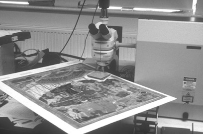
Figure 3. The Raman microscope in use. Photography by Mike Wheeler. (click image for larger version)
Building on previous information about pigments from three of the folios, which had been obtained by optical microscopy and UV examination, Raman spectroscopy was used to examine the pigments. Between thirty and forty sites from each painting were analysed. These were selected to provide as full a representation of the complete palette as possible. The speed and ease with which this number of readings could be taken with the Raman microscope, when compared to other analytical techniques, was a great advantage.
Building on previous information about pigments from three of the folios, which had been obtained by optical microscopy and UV examination, Raman spectroscopy was used to examine the pigments. Between thirty and forty sites from each painting were analysed. These were selected to provide as full a representation of the complete palette as possible. The speed and ease with which this number of readings could be taken with the Raman microscope, when compared to other analytical techniques, was a great advantage.
Conclusion
The application of Raman microscopy is particularly useful in paper conservation where analysis often has to be carried out on thin layers of materials and sampling is not an option. In the above examples, the non-destructive, non-invasive nature of Raman microscopy was paramount in selecting it as an analytical tool. Other advantages include the portability of the instrument, which facilitates on site analysis and the relative speed at which the analysis can be carried out allowing a large number of readings to be taken in a short time.
Acknowledgements:
Sigrun Thiel; Katherine Brown and Greg Smith, Chemistry Department, UCL; Dr Robert Withnal, Chemistry and Life Sciences, University of Greenwich
Spring 2002 Issue 40
- Editorial
- Sustainability and precaution - Part 1
- People watching: Monitoring heritage hospitality functions in historic houses
- Ethical considerations in the treatment of a Tibetan sculpture
- Building models: Comparative swelling powers of organic solvents on oil paint and the cleaning of paintings
- Further applications of Raman microscopy in paper conservation
- Critical evaluation of laser cleaning of parchment documents
- Collaboration
- Printer friendly version